Tadalafil entfaltet seine Wirkung über eine selektive Hemmung der PDE5, wodurch die Konzentration von cGMP im glatten Muskelgewebe stabil bleibt. Diese biochemische Modulation resultiert in einer langanhaltenden Relaxation der Gefäßwände. Der Wirkstoff wird nach oraler Einnahme effizient resorbiert, mit einer Bioverfügbarkeit von rund 80 %. Seine Halbwertszeit von bis zu 36 Stunden ist innerhalb dieser Substanzklasse außergewöhnlich. Abgebaut wird er in der Leber, hauptsächlich durch CYP3A4, mit anschließender biliärer Exkretion. Typische unerwünschte Wirkungen entstehen durch eine verstärkte Vasodilatation, etwa Kopfschmerzen oder Flush. Pharmakologisch wird cialis generika vor allem durch die verlängerte Wirkungsdauer charakterisiert.
Doi:10.1016/j.cimid.2004.01.005
& Infectious Diseases 27 (2004) 413–422
Acute phase response in reindeer after challenge
T. Orroa,*, S. Sankaria, T. Pudasb, A. Oksanenc, T. Soveria
aFaculty of Veterinary Medicine, Department of Clinical Veterinary Sciences, University of Helsinki,
bDepartment of Biology, University of Oulu, P.O. BOX 333, 90571 Oulu, Finland
cNational Veterinary and Food Research Institute, EELA, P.O. BOX 517, 60101, Oulu, Finland
The serum concentrations of two acute phase proteins (APPs), haptoglobin (Hp) and serum
amyloid-A (SAA), were monitored in reindeer after challenge with endotoxin. Four adult femalereindeer received either 0.1 mg/kg Escherichia coli 0111:B4 lipopolysaccharide B or saline solutionintravenously. At the second challenge, the treatments were reversed. In addition to the APPs,changes in blood chemistry and rectal temperature were monitored. The endotoxin challenge causeda significant increase in SAA (peak 48 h) and a sharp decrease (8 – 12 h) of serum iron concentrationsin all animals. The mean Hp concentration increased at 8 h and remained elevated until 48 h, but nostatistically significant differences were found. This investigation demonstrates that challenge with asingle-bolus dose of E. coli endotoxin can activate the acute phase response (APR) and SAA appearsto be a more sensitive indicator of the APR than Hp during bacterial infection in reindeer.
q 2004 Elsevier Ltd. All rights reserved.
Keywords: Acute phase response (APR); Endotoxin; Haptoglobin (Hp); Reindeer; Serum amyloid-A (SAA)
Les concentrations de deux prote´ines de l’inflammation, l’haptoglobine (Hp) et l’amyloı¨de A
se´rique (SAA), la tempe´rature rectale et les parame`tres biochimiques sanguins ont e´te´ suivis chezquatre rennes adultes femelles ayant rec¸u lors d’une premie`re inoculation intraveineuse soit0.1 mg/kg de lipopolysaccharide B d’Escherichia coli O111:B4, soit du se´rum physiologique, puislors d’une seconde inoculation l’inverse.
L’injection d’endotoxine a entraıˆne´ une augmentation significative de la concentration de SAA
(pic a` 48 h) et une diminution brutale (8 – 12 h) du fer se´rique chez tous les animaux. La concentration
* Corresponding author. Tel.: þ358-19-529-5304; fax: þ358-19-685-1181.
E-mail address: toomas.orro@helsinki.fi (T. Orro).
0147-9571/$ - see front matter q 2004 Elsevier Ltd. All rights reserved. doi:10.1016/j.cimid.2004.01.005
T. Orro et al. / Comp. Immun. Microbiol. Infect. Dis. 27 (2004) 413–422
moyenne d’Hp a augmente´ a` 8 h et est reste´e e´leve´e jusqu’a` 48 h, mais sans qu’aucune diffe´rencestatistiquement significative ne soit trouve´e. Ceci de´montre qu’une inoculation unique d’endotoxined’E. coli peut de´clencher une re´action inflammatoire chez le renne et qu’en cas d’infectionbacte´rienne la SAA apparaıˆt eˆtre plus sensible que l’Hp pour de´tecter cette re´action.
q 2004 Elsevier Ltd. All rights reserved.
Mots-cle´: Re´action inflammatoire; Endotoxine; Haptoglobine; Amyloı¨de A se´rique; Renne
The inflammatory reaction is a series of complex physiological events occurring in the
host after tissue injury or infection. The purposes of these events are to eliminate theinfecting agent, prevent further tissue damage and restore the homeostasis of the hostorganism. The early sets of reactions that occur immediately after tissue damage are knownas the acute phase response (APR). It is characterized by the presence of an inflammatoryreaction at the site of injury or infection and systemically by multiple changes throughoutthe organism such as fever, depression, leucocytosis, increased permeability of bloodvessels, stimulation of adrenocorticotropic hormone (ACTH) production, redistribution oftrace elements, etc. One of the main changes occurring during the APR is hepatic productionof various plasma proteins These so-called acute phase proteins (APPs) play a veryimportant role in the defense response of the host. For example, some APPs are involved innonspecific protection against infections caused by gram-negative bacteria , but in manycases their exact physiological function is not clear.
Monitoring the plasma concentrations of APPs can provide information on progression
of the APR. Their potential use in veterinary medicine has been extensively investigatedand they are already widely used as markers of disease in veterinary and medical science. Some of the many applications for APPs include evaluation of collective health of herdsby detection of subclinical diseases and identification of animals with inflammatorylesions at slaughter. Thus, APPs may also serve as valuable tools in reindeer husbandry aswell as in giving diagnostic information on the detection, prognosis and monitoring ofdiseases in reindeer.
Although the APR is highly conserved in nature, APP profiles show significant variability
between different species Haptoglobin (Hp) has been the APP most commonly monitoredas a marker for inflammation in cattle and other domestic ruminants. The plasma Hpconcentration is very low in healthy bovines but during the APR it can increase over 100-fold. Serum amyloid-A (SAA) is another major APP in many animal species, including cattle,and has been shown to be a good diagnostic marker for determining the presence of infectionsand inflammatory conditions Evaluation of the concentrations of different APPsallows better estimates of the complex systemic effects of inflammatory mediators duringvarious inflammatory conditions, e.g. in distinguishing between acute and chronicinflammation and for evaluation of disease severity.
The main objective here was to investigate the host response of reindeer (Rangifer
tarandus tarandus) after bacterial endotoxin challenge as an acute phase stimulant and toevaluate the changes occurring in serum concentrations of SAA and Hp as potential
T. Orro et al. / Comp. Immun. Microbiol. Infect. Dis. 27 (2004) 413–422
markers of infections. In addition to the APPs, other changes occurring in blood chemistryand in some clinical parameters were also monitored.
This experiment was carried out at the Zoological Gardens of the Department of
Biology, University of Oulu, Finland. The first experiment was initiated on February 18,2002 and the second on April 8, 2002. Eight adult female reindeer were randomly dividedinto two equal groups. The mean weight of the animals was 76.7 kg (range 67.5 – 90.5 kg)and age was 5 years (3 – 6 years). The groups were kept in two separate corrals (about650 m2 each) in which they underwent a long adaptation period before the experiment wasinitiated. The animals were fed with lichen (Cladina spp.), commercial reindeer pelletsand fresh water ad libitum. The first group received 0.1 mg/kg E. coli 0111:B4lipopolysaccharide B ((LPS); 1 mg/ml Bactoq, Difco Laboratories, Inc., Detroit, MI,USA) into the jugular vein. The second group received the same volume of physiologicalsodium chloride solution (0.1 ml/kg). After 7 weeks, the same procedure was repeated viceversa. The dose was chosen based on published studies in small domestic ruminants suchas goat In these experiments, this endotoxin dose resulted in clear activation ofthe APR, as shown by significant decrease in serum iron concentrations and increase inrectal temperature and serum cortisol concentrations. This dose caused only modest andshort-acting general signs of disease, e.g. depression and alteration of rumen contractility. The rectal temperature was recorded at the same time that blood was taken during the firstday of the experiments. Clinical observations without manual handling were done everyhour during the first 12 h. After the experiments, the animals were killed and autopsiesperformed.
The group of reindeer was herded into the small pen near the corrals and then caught
and manually restrained. Blood samples were taken from the jugular vein before theendotoxin or physiological saline solution injections and after 1, 4, 8, 12, 24, 48, 96 and168 h into plain tubes. The serum was separated, frozen in portions and stored at 2 20 8Cfor further analysis.
Serum Hp was determined using the hemoglobin-binding assay described by Makimura
and Suzuki with slight modifications, in which tetramethylbenzidine (0.06 mg/ml) wasused as substrate SAA was measured with a commercially available ELISA kit (PhaseSAA kit, Tridelta Ltd, Ireland), according to the manufacturer’s instructions for cattle.
The activities of serum aspartate aminotransferase (ASAT) and creatine kinase (CK)
were determined following the recommendations of the Scandinavian Society for Clinical
T. Orro et al. / Comp. Immun. Microbiol. Infect. Dis. 27 (2004) 413–422
Chemistry and Clinical Physiology Spectrophotometric methods were used forthe determination of serum total protein , urea sorbitol dehydrogenase (SDH)albumin and g-glutamyl transferase (GGT) according to the InternationalFederation of Clinical Chemistry and Labarotory Medicine (IFCC) Expert Panel onEnzymes . Serum iron was determined with a colorimetric method . The analyseswere performed with an automatic chemistry analyzer (KONE Pro, Konelab, ThermoClinical Labsystems Oy, Vantaa, Finland). Cortisol was measured in 25-mL duplicatesusing radioimmunoassay with Coat-A-Count RIA kits obtained from Diagnostic ProductsCorporation (Los Angeles, CA, USA). All results are expressed as mean ^ standarddeviation (SD).
Carry-over and period effects were tested using the t-test procedures of Jones and
Kenward These tests were run for selected time points, in cases of insignificant carry-over effects in the examined parameters, the data from both challenges were combined. Differences between the E. coli LPS treatment and control groups were analyzed usingrepeated measures analyses of variance with treatment and time after challenge as withinfactors. Greenhouse – Geiser adjusted p-values were used for evaluation of results, whichwere significant at p , 0:05:
After laboratory analysis, two reindeer were excluded from statistical analysis, one
from each group. One reindeer already showed increased Hp (over 3-fold) and SAA (over10-fold) concentrations in the 0-h sample, which remained high during the firstexperiment. Clear arthritic changes were found in the left tarsal joint of this animal atautopsy; in all other reindeer no pathological changes were found. The second excludedreindeer reacted very strongly to the restraint procedures used at the time of bloodsampling; its serum CK and ASAT activities increased markedly during the first 24 h ofexperiments, indicating muscle injury.
A carry-over effect was found in cortisol and data analysis was carried out separately in
both experiments for that variable. Period differences could be seen in iron ðp , 0:05Þ andin the activities of ASAT ðp , 0:01Þ and GT ðp , 0:05Þ: In the first experiment, allanimals receiving LPS showed clear signs of general depression (they kept their headsdown and moved slowly) and two had tremor of the legs. These clinical signs wereobserved during the first 4 h after LPS administration. The same reindeer had loosedroppings on the following day. The signs of depression were less obvious during thesecond experiment.
Endotoxin administration caused a significant increase in the concentration of SAA
(p , 0:001Þ: The rise in SAA in the LPS group was seen in the 12-h sample andpeaked at 48 h ð184:7 ^ 89:4 mg=lÞ: At this time, the LPS-treated animals displayed a 2 to29-fold increase of serum SAA from the baseline level. The mean SAA concentrations inthe 96-h sample were decreased below the pretreatment levels in both groups.
T. Orro et al. / Comp. Immun. Microbiol. Infect. Dis. 27 (2004) 413–422
Fig. 1. Mean (^ SD) serum concentrations of SAA (a), Hp (b) and iron (c) in reindeer ðn ¼ 6Þ after IVadministration of E. coli LPS (S; 0.1 mg/kg) or saline (V) at 0 h. The data are combined from two successivechallenges. Statistical differences are given in the text.
The mean serum Hp concentrations showed a small tendency to decrease during the
first 4-h period in the LPS group, increased slightly in the 8-h sample and attainedmaximum value at 24 h (0.87 ^ 0.25 g/l). The Hp concentrations remained relativelystable in the control group throughout the experiment (); however, these differencesbetween treatments were not statistically significant. Wide individual variation occurred inHp response to endotoxin administration; only one reindeer showed a near 2-fold increasein the 48-h sample and one showed no increase at any time.
Serum iron concentrations decreased sharply in all endotoxin-treated animals and were
significantly lower ðp , 0:01Þ at 8 and 12 h (). The serum cortisol concentrationsshowed wide variability among animals, but no consistent increases in cortisol in the LPS
T. Orro et al. / Comp. Immun. Microbiol. Infect. Dis. 27 (2004) 413–422
group were detected during the follow-up period ð0 – 96 hÞ at the time of both sets of theexperiment. The only exception was at the time of the first experiment when the LPS groupshowed values clearly elevated over those of the control animals in the 4-h sample. Thereindeer already had showed very high cortisol values in the pretreatment sample (mean102.9 nmol/l, range 9.8 – 240 nmol/l;
Serum urea values began to increase after treatment in both groups simultaneously and
after 4 h decreased slowly until 96 h to under the baseline level (time effectp , 0:001Þ: After one week, the mean urea concentration had nearly returned to thepretreatment level. There were no differences between treatment groups at any time point.
Fig. 2. Mean (^ SD) serum concentrations of urea (a) n ¼ 6; cortisol from first set (b) n ¼ 3 and from second setof the experiment (c) n ¼ 3 in reindeer after IV administration of E. coli LPS (S; 0.1 mg/kg) or saline (V) at 0 h. The urea data are combined from two successive challenges. Statistical differences are given in the text.
T. Orro et al. / Comp. Immun. Microbiol. Infect. Dis. 27 (2004) 413–422
Mean rectal temperatures were already higher before treatment in the LPS group and
decreased steadily in both groups throughout the 24-h period, which was recorded, but thisdifference was not significant. The changes in concentrations of serum total protein,albumin, ASAT, CK, GT and SDH recorded during the follow-up period werenonsignificant between the endotoxin and control groups.
Bacterial endotoxins, which are LPSs from the cell wall of gram-negative bacteria, are
considered to cause most pathophysiological reactions during bacterial infections. Thephysiological effects of LPS are based predominantly on activation of various molecularmediators such as the cytokines tumor necrosis factor (TNF)-a, interleukin (IL)-1band IL-6, which are released in response to LPS predominantly by monocytes andmacrophages These inflammatory mediators initiate host inflammatory response andthe production of hepatic APPs. Since cytokines are the main stimulators of excretion ofAPPs from the liver , LPS administration has been used in APP research as an acutephase stimulant in bovines In the present study, injection of E. coli LPS inducedactivation of cytokine release in reindeer as shown by significant decrease in serum ironconcentration and increase in serum SAA concentrations in all LPS-challenged animals. These changes were similar to those previously reported in domestic ruminants . Aslight tendency for Hp concentrations to increase was seen earlier than SAA followingLPS challenge, but the Hp patterns differed widely between individuals and the relativeincreases were small. A similar, evident but nonsignificant elevation of Hp was reportedpreviously in pigs after a single-bolus LPS dose administration but the Hp responsewas clearly elevated when repeated injections were used Hp and SAA differ in theircytokine-controlled induction in the liver, and generally Hp needs stronger cytokinestimulation to be produced than SAA The cytokine response sequence is notaffected by LPS dose or administration route, but the amplitude of the response is dosedependent and a single-bolus dose of E. coli LPS may have been insufficient to initiatethe cytokine secretion needed for prolonged Hp increase. This is supported by other studiesin bovines in which Hp increased after experimental gram-negative infections when stronger and prolonged cytokine release occurs, or after higher E. coli LPS doseadministration
Variability in APP expression during the APR between animal species can also explain
the low Hp response observed. However, the serum Hp concentration can increase many-fold over the baseline level and appears to act as an APP in reindeer. This was observed inthe reindeer we had to remove from our study due to the presence of exceptionally highSAA and Hp concentrations probably resulting from arthritis.
Another explanation for the mild APR to endotoxaemia may be the stress reactions of
reindeer. Although treatment did not affect the urea concentration, increased concen-trations occurring during the first blood samplings and subsequent decrease when animalsbecame used to handling, further confirm the presence of a strong stress response inreindeer. Increased concentrations of serum urea in stressed reindeer have been reportedand significantly higher urea concentrations have been seen in intensively handled
T. Orro et al. / Comp. Immun. Microbiol. Infect. Dis. 27 (2004) 413–422
reindeer versus animals subjected to minimal handling The presence of a carry-overeffect in the cortisol results is very difficult to explain, especially when considering that wehad long wash-out period between experiment sets. However, the similarities in meancortisol concentration patterns within the animal groups during both experiments,regardless of LPS challenge and wide individual variability (), indicate that cortisolwas more affected by the experimental procedure than by the presence of endotoxaemiaand that the carry-over effect was at least partly the result of stress responses of individualanimals to handling. The elevated cortisol levels observed before LPS challenge in thisstudy probably affected the results. High endogenous glucocorticosteroid concentrationscan influence the APR and expression of APPs in many ways; e.g. it can directly stimulateAPP production in the liver, but this is not believed to be a very powerful response andadministration of dexamethasone failed to stimulate APP in cows . On the other hand,cortisol can inhibit APP increase by decreasing the cytokine response. Endogenous andexogenous glucocorticoids can suppress TNF-a production after endotoxin administrationand therefore the stress reaction induced by handling before endotoxinadministration may have an inhibiting effect on proinflammatory cytokine release andmay be one reason for the mild Hp response to the endotoxin dose used in this study. Further support for the inhibitory effect of stress reactions before LPS administration isgiven by the fact that no fever response occurred, although the clinical, serum iron andSAA responses were clearly present. Cortisol and fever responses occurring duringendotoxaemia to some extent share a common mediating factor, namely prostaglandin,which is mediated by proinflammatory cytokines and the controlling mechanisms forfever and changes in serum iron concentrations are independent
In conclusion, we have demonstrated that single-dose E. coli endotoxin challenge
activates the APR and that this model can be used in reindeer for examining thepathophysiology of inflammation and infection, although higher doses may be moreappropriate. However, in semidomesticated animals such as reindeer, stress reactions fromhandling are very strong and their possible effects on the results must be addressed. SAAappears to be a more sensitive indicator of the APR than Hp in reindeer after LPSchallenge. Since these proteins have different stimulation patterns, monitoring of bothproteins can provide more information on the ongoing APR of the host and thus mayprovide a useful tool in veterinary medical science in reindeer. Further investigation isneeded to increase our understanding of the expression of these proteins in cases of variousnaturally occurring and experimental infections or inflammations.
[1] Baumann H, Gauldie J. The acute phase response. Immunol Today 1994;15:74 – 80. [2] Hochepied T, Van Molle W, Berger FG, Baumann H, Libert C. Involvement of the acute phase protein alpha
1-acid glycoprotein in non-specific resistance to a lethal gram-negative infection. J Biol Chem 2000;275:14903– 9.
[3] Kushner I, Mackiewicz A. Acute phase proteins as disease markers. Dis Markers 1987;5:1– 11. [4] Eckersall PD, Conner JG. Bovine and canine acute phase proteins. Vet Res Commun 1988;12:169 – 78. [5] Conner JG, Eckersall PD, Wiseman A, Bain RK, Douglas TA. Acute phase response in calves following
infection with Pasteurella haemolytica, Ostertagia ostertagi and endotoxin administration. Res Vet Sci1989;47:203 – 7.
T. Orro et al. / Comp. Immun. Microbiol. Infect. Dis. 27 (2004) 413–422
[6] Skinner JG, Roberts L. Haptoglobin as an indicator of infection in sheep. Vet Rec 1994;134:33– 6. [7] Alsemgeest SPM, Kalsbeek HC, Wensing T, Koeman JP, van Ederen AM, Gruys E. Concentrations of
serum amyloid-A (SAA) and haptoglobin (Hp) as parameters of inflammatory diseases in cattle. Vet Quart1994;16:21 – 3.
[8] Wittum TE, Young CR, Stanker LH, Griffin DD, Perino LJ, Littledike ET. Haptoglobin response to clinical
respiratory tract disease in feedlot cattle. Am J Vet Res 1996;57:646 – 9.
[9] Horadagoda A, Eckersall PD, Alsemgeest SPM, Gibbs HA. Purification and quantitative measurement of
bovine serum amyloid-A. Res Vet Sci 1993;55:317– 25.
[10] Heegaard PM, Godson DL, Toussaint MJ, Toornehooj K, Larsen LE, Viuff B, Roonsholt L. The acute phase
response of haptoglobin and serum amyloid A (SAA) in cattle undergoing experimental infection withbovine respiratory syncytial virus. Vet Immunol Immunopathol 2000;77:151– 9.
[11] Eckersall PD, Young FJ, McComb C, Hogarth CJ, Safi S, Weber A, McDonald T, Nolan AM, Fitzpatrick JL.
Acute phase proteins in serum and milk from dairy cows with clinical mastitis. Vet Rec 2001;148:35– 41.
[12] van Miert AS, van Duin CT, Verheijden JH, Schotman AJ. Staphylococcal enterotoxin B and Escherichia
coli endotoxin: comparative observations in goats on fever and associated clinical hematologic and bloodbiochemical changes after intravenous and intramammary administration. Am J Vet Res 1983;44:955– 63.
[13] van Miert AS, van Duin CT, Wensing T. Fever and acute phase response induced in dwarf goats by
endotoxin and bovine and human recombinant tumour necrosis factor alpha. J Vet Pharmacol Ther 1992;15:332 – 42.
[14] Massart-Leen AM, Burvenich C, Vandeputte-Van Messom G, Hilderson H. Partial prostaglandin-mediated
mechanism controlling the release of cortisol in plasma after intravenous administration of endotoxins. Domest Anim Endocrinol 1992;9:273 – 83.
[15] Makimura S, Suzuki N. Quantitative determination of bovine serum haptoglobin and its elevation in some
inflammatory diseases. Jpn J Vet Sci 1982;44:15– 21.
[16] Scandinavian Society for Clinical Chemistry and Clinical Physiology, Recommended methods for the
determination of four enzymes in blood. Scan J Clin Lab Investig 1974;33:291– 306.
[17] Scandinavian Society for Clinical Chemistry and Clinical Physiology, Recommended methods for the
determination of creatine kinase in blood modified by the inclusion of EDTA. Scan J Clin Lab Investig1979;39:1– 5.
[18] Weichselbaum TE. An accurate and rapid method for the determination of proteins in small amounts of
blood serum and plasma. Am J Clin Pathol 1946;16:40 – 9.
[19] Gutmann I, Bergmeyer HU. In: Bergmeyer HU, editor. Determination of urea with glutamate
dehydrogenase as indicator enzyme, 2nd English ed. Methods of enzymatic analysis, vol. 4. New York:Academic Press; 1974. p. 1794 – 8.
[20] Gerlach U, Hiby W. Sorbitol dehydrogenase. In: Bergmeyer HU, editor. Methods of enzymatic analysis,
vol. 2. New York: Academic Press; 1974. p. 569 – 73.
[21] Doumas BT, Watson WA, Biggs H. Albumin standards and the measurement of serum albumin with
bromcresol green. Clin Chim Acta 1971;31:87– 96.
[22] IFCC Expert Panel on Enzymes, IFCC methods for the measurement of the catalytic concentration of
enzymes. Part 4. IFCC method for g-glutamyl transferase. J Clin Chem Clin Biochem 1983;21:633 – 46.
[23] Persijn J-P, Van der Slik W, Reithorst A. Determination of serum iron and latent iron-binding capacity
(LIBC). Clin Chim Acta 1971;35:91– 8.
[24] Jones B, Kenward MG. Design and analysis of crossover trials. London: Chapmann & Hall; 1989. [25] Lohuis JACM, Verheijden JHM, Burvenich C, van Miert ASJPAM. Pathophysiological effects of
endotoxins in ruminants. 1. Changes in body temperature and reticulo-rumen motility, and the effects ofrepeated administration. Vet Quart 1988;10:109– 16.
[26] Henderson B, Wilson M. Cytokine induction by bacteria: beyond lipopolysaccharide. Cytokine 1996;8:
[27] Boosman R, Niewold ThA, Mutsaers CWAAM, Gruys E. Serum amyloid A concentrations in cows given
endotoxin as an acute-phase stimulant. Am J Vet Res 1989;50:1690– 4.
[28] Werling D, Sutter F, Arnold M, Kun G, Tooten PCJ, Gruys E, Kreuzer M, Langhans W. Characterisation of
the acute phase response of heifers to a prolonged low dose infusion of lipopolysaccharide. Res Vet Sci1996;61:252 – 7.
T. Orro et al. / Comp. Immun. Microbiol. Infect. Dis. 27 (2004) 413–422
[29] Van Miert ASJPAM, Van Duin CTM, Wensing Th. Fever and changes in plasma zinc and iron
concentrations in the goat. The effects of interferon inducers and recombinant IFN-alpha 2a. J Comp Pathol1990;103:289 – 300.
[30] Wright KJ, Balaji R, Hill CM, Dritz SS, Knoppel EL, Minton JE. Integrated adrenal, somatotropic, and
immune responses of growing pigs to treatment with lipopolysaccharide. J Anim Sci 2000;78:1892– 9.
[31] Dritz SS, Owen KQ, Goodband RD, Nelssen JL, Tokach MD, Chengappa MM, Blecha F. Influence of
lipopolysaccharide-induced immune challenge and diet complexity on growth performance and acute-phaseprotein production in segregated early-weaned pigs. J Anim Sci 1996;74:1620 – 8.
[32] Alsemgeest SPM, van’t Klooster GAE, van Miert ASJPAM, Hulskamp-Koch CK, Gruys E. Primary bovine
hepatocytes in the study of cytokine induced acute-phase protein secretion in vitro. Vet ImmunolImmunopathol 1996;53:179 – 84.
[33] Gerros TC, Semrad SD, Proctor RA, LaBorde A. Effect of dose and method of administration of endotoxin
on cell mediator release in neonatal calves. Am J Vet Res 1993;54:2121 – 7.
[34] Godson DL, Campos M, Attah-Poku SK, Redmond MJ, Cordeiro DM, Sethi MS, Harland RJ, Babiuk LA.
Serum haptoglobin as an indicator of the acute phase response in bovine respiratory disease. Vet ImmunolImmunopathol 1996;51:277 – 92.
[35] Horadagoda NU, Hodgson JC, Moon GM, Wijewardana TG, Eckersall PD. Role of endotoxin in the
pathogenesis of haemorrhagic septicaemia in the buffalo. Microb Pathog 2001;30:171– 8.
[36] Hyva¨rinen H, Helle T, Nieminen M, Va¨yrynen P, Va¨yrynen R. Some effects of handling reindeer during
gathering on the composition of their blood. Anim Prod 1976;22:105 – 14.
[37] Rehbinder C, Edqvist LE. Influence of stress on some blood constituents in reindeer (Rangifer tarandus L).
[38] Wiklund E, Rehbinder C, Malmfors G, Hansson I, Danielsson-Tham M-L. Ultimate pH values and
bacteriological condition of meat and stress metabolites in blood of transported reindeer bulls. Rangifer2001;21:3– 12.
[39] van der Kolk JH, Alsemgeest SP, Wensing T, Niewold TA, Gruys E, Mol JA, Breukink HJ. Failure of
adrenocorticotrophic hormone to release serum amyloid A in cattle. Res Vet Sci 1992;52:113 – 4.
[40] Zuckerman SH, Shellhaas J, Butler LD. Differential regulation of lipopolysaccharide-induced interleukin-1
and tumor necrosis factor synthesis: effects of endogenous and exogenous glucocorticoids and the role of thepituitary-adrenal axis. Eur J Immunol 1989;19:301– 5.
O elixir da longa vida Honoré de Balzac Nos começos da vida literária do autor, um seu amigo, morto há muito tempo, deu-lhe o assunto para este estudo, que mais tarde encontrou numa colecção publicada nos princípios deste século. Segundo as suas conjecturas, trata-se de uma fantasia devida a um tal Hoffman, de Berlim, publicada nalgum almanaque alemão e esquecida pelos edito
Nome programma:Nucleic acids as drugs and drug targets: development of oligonucleotide-based drugsand characterization of the mechanism of action of nucleic acid- and kinase-targetedcompoundsObiettivi specifici del programmaThe goals of this program focuses on the following lines of research:1. The development of nucleic acids as drugs employing the technique known asSELEX, or Systematic Evolut


 & Infectious Diseases 27 (2004) 413–422
Acute phase response in reindeer after challenge
T. Orroa,*, S. Sankaria, T. Pudasb, A. Oksanenc, T. Soveria
aFaculty of Veterinary Medicine, Department of Clinical Veterinary Sciences, University of Helsinki,
bDepartment of Biology, University of Oulu, P.O. BOX 333, 90571 Oulu, Finland
cNational Veterinary and Food Research Institute, EELA, P.O. BOX 517, 60101, Oulu, Finland
The serum concentrations of two acute phase proteins (APPs), haptoglobin (Hp) and serum
amyloid-A (SAA), were monitored in reindeer after challenge with endotoxin. Four adult femalereindeer received either 0.1 mg/kg Escherichia coli 0111:B4 lipopolysaccharide B or saline solutionintravenously. At the second challenge, the treatments were reversed. In addition to the APPs,changes in blood chemistry and rectal temperature were monitored. The endotoxin challenge causeda significant increase in SAA (peak 48 h) and a sharp decrease (8 – 12 h) of serum iron concentrationsin all animals. The mean Hp concentration increased at 8 h and remained elevated until 48 h, but nostatistically significant differences were found. This investigation demonstrates that challenge with asingle-bolus dose of E. coli endotoxin can activate the acute phase response (APR) and SAA appearsto be a more sensitive indicator of the APR than Hp during bacterial infection in reindeer.
& Infectious Diseases 27 (2004) 413–422
Acute phase response in reindeer after challenge
T. Orroa,*, S. Sankaria, T. Pudasb, A. Oksanenc, T. Soveria
aFaculty of Veterinary Medicine, Department of Clinical Veterinary Sciences, University of Helsinki,
bDepartment of Biology, University of Oulu, P.O. BOX 333, 90571 Oulu, Finland
cNational Veterinary and Food Research Institute, EELA, P.O. BOX 517, 60101, Oulu, Finland
The serum concentrations of two acute phase proteins (APPs), haptoglobin (Hp) and serum
amyloid-A (SAA), were monitored in reindeer after challenge with endotoxin. Four adult femalereindeer received either 0.1 mg/kg Escherichia coli 0111:B4 lipopolysaccharide B or saline solutionintravenously. At the second challenge, the treatments were reversed. In addition to the APPs,changes in blood chemistry and rectal temperature were monitored. The endotoxin challenge causeda significant increase in SAA (peak 48 h) and a sharp decrease (8 – 12 h) of serum iron concentrationsin all animals. The mean Hp concentration increased at 8 h and remained elevated until 48 h, but nostatistically significant differences were found. This investigation demonstrates that challenge with asingle-bolus dose of E. coli endotoxin can activate the acute phase response (APR) and SAA appearsto be a more sensitive indicator of the APR than Hp during bacterial infection in reindeer.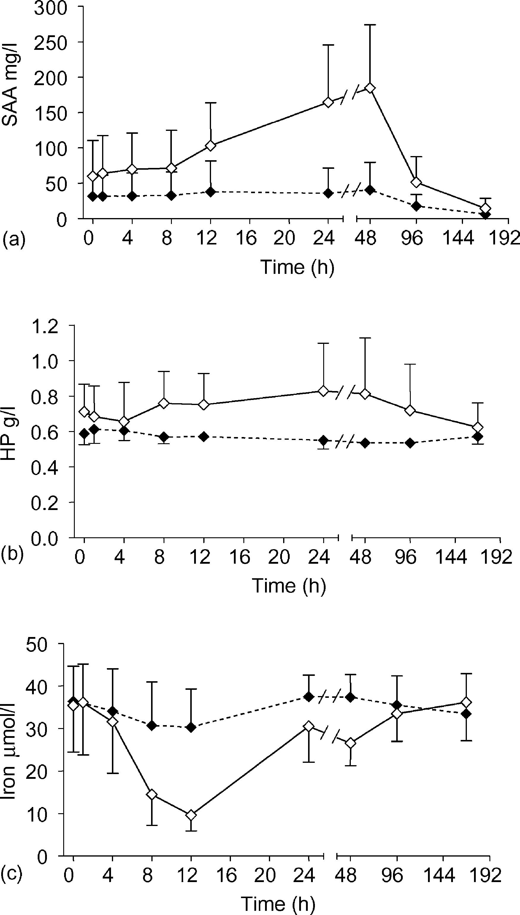 T. Orro et al. / Comp. Immun. Microbiol. Infect. Dis. 27 (2004) 413–422
Fig. 1. Mean (^ SD) serum concentrations of SAA (a), Hp (b) and iron (c) in reindeer ðn ¼ 6Þ after IVadministration of E. coli LPS (S; 0.1 mg/kg) or saline (V) at 0 h. The data are combined from two successivechallenges. Statistical differences are given in the text.
T. Orro et al. / Comp. Immun. Microbiol. Infect. Dis. 27 (2004) 413–422
Fig. 1. Mean (^ SD) serum concentrations of SAA (a), Hp (b) and iron (c) in reindeer ðn ¼ 6Þ after IVadministration of E. coli LPS (S; 0.1 mg/kg) or saline (V) at 0 h. The data are combined from two successivechallenges. Statistical differences are given in the text.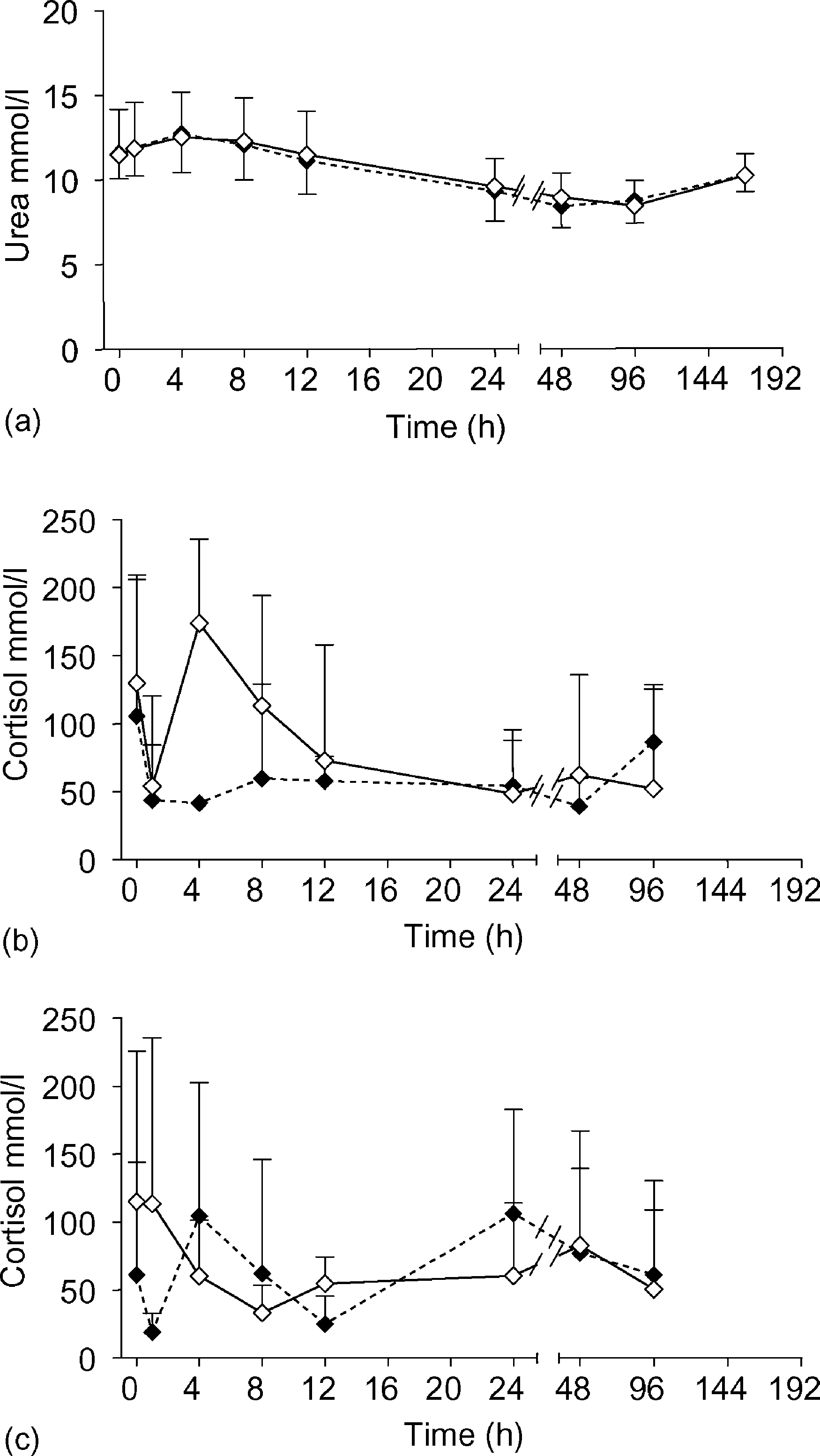 T. Orro et al. / Comp. Immun. Microbiol. Infect. Dis. 27 (2004) 413–422
group were detected during the follow-up period ð0 – 96 hÞ at the time of both sets of theexperiment. The only exception was at the time of the first experiment when the LPS groupshowed values clearly elevated over those of the control animals in the 4-h sample. Thereindeer already had showed very high cortisol values in the pretreatment sample (mean102.9 nmol/l, range 9.8 – 240 nmol/l;
Serum urea values began to increase after treatment in both groups simultaneously and
after 4 h decreased slowly until 96 h to under the baseline level (time effectp , 0:001Þ: After one week, the mean urea concentration had nearly returned to thepretreatment level. There were no differences between treatment groups at any time point.
T. Orro et al. / Comp. Immun. Microbiol. Infect. Dis. 27 (2004) 413–422
group were detected during the follow-up period ð0 – 96 hÞ at the time of both sets of theexperiment. The only exception was at the time of the first experiment when the LPS groupshowed values clearly elevated over those of the control animals in the 4-h sample. Thereindeer already had showed very high cortisol values in the pretreatment sample (mean102.9 nmol/l, range 9.8 – 240 nmol/l;
Serum urea values began to increase after treatment in both groups simultaneously and
after 4 h decreased slowly until 96 h to under the baseline level (time effectp , 0:001Þ: After one week, the mean urea concentration had nearly returned to thepretreatment level. There were no differences between treatment groups at any time point.