Tadalafil entfaltet seine Wirkung über eine selektive Hemmung der PDE5, wodurch die Konzentration von cGMP im glatten Muskelgewebe stabil bleibt. Diese biochemische Modulation resultiert in einer langanhaltenden Relaxation der Gefäßwände. Der Wirkstoff wird nach oraler Einnahme effizient resorbiert, mit einer Bioverfügbarkeit von rund 80 %. Seine Halbwertszeit von bis zu 36 Stunden ist innerhalb dieser Substanzklasse außergewöhnlich. Abgebaut wird er in der Leber, hauptsächlich durch CYP3A4, mit anschließender biliärer Exkretion. Typische unerwünschte Wirkungen entstehen durch eine verstärkte Vasodilatation, etwa Kopfschmerzen oder Flush. Pharmakologisch wird cialis generika vor allem durch die verlängerte Wirkungsdauer charakterisiert.
[pdf] in cell analyzer 3000 high throughput multiplexed cellular toxicity
High Throughput Multiplexed Cellular Toxicity
*Jan Turner, Samantha Murphy, Elaine Adie, Angela Williams, Molly Price-Jones. Amersham Biosciences Limited, Amersham Place, Little Chalfont, Buckinghamshire, HP7 9NA, England., email: jan.turner@uk.amershambiosciences.com.
Alamar Blue was obtained from Serotec , UK, cells were
plus the cell count were obtained from each well as an
Conventional cellular toxicological assays are based on
incubated for 4 hours and fluorescence was measured
homogenous assay. The Alamar Blue results showed
the microscopic observation of toxic end points in fixed
no significant change with dose (Fig. 2).
cells and for these reasons, are inherently limited both
in terms of data throughput and content. Using
Following the exposure period, cells were aspirated and
commercially available apoptotic markers and the IN
washed briefly with 100µl warmed phosphate buffered
Cell Analyzer 3000, we have demonstrated and
saline (PBS). This was followed by incubation with
quantified multiple sub-toxic responses in individual
100µl of probe mixture for a total of 15 min 37 C before
cells in both live and fixed heterogeneous primary cell
being read using the IN Cell Analyzer 3000 (Fig. 1 and
cultures and immortalized cell lines, while processing
up to 40,000 data points in one plate. Pre-apoptotic
perturbances have been simultaneously quantitated in
Following Staurosporine exposure, Hoechst 33342 (Fig.
metabolically competent rat hepatocytes, enabling
1A & B), Mitotracker (Fig. 1A & C) and histology
mechanistic and pathway analyses to be performed. In
staining (Fig.1A & D) showed measureable changes. Fig.4. Multiple homogeneous assay readouts following the exposure
addition, correlation between conventional staining,
Hoechst showed statistically significant increases in
of primary rat hepatocytes to Staurosporine for 4 hours: comparison to
morphological changes and sub toxic responses have
punctate staining of the nucleus with increases in dose. Alamar Blue assay. Values are means +/- 1 SD of n=16.
been clearly demonstrated using the IN Cell Analyzer.
Mitotracker showed a decrease in the characteristic
punctate stained cytoplasm with increasing dose. The
Fig. 2. Multiple homogeneous assay readouts following theMitotracker
P450 reductase histological stain showed a quantitative
exposure of primary rat hepatocytes to Staurosporine for 4 hours:
increase in the intensity of staining in the cytoplasm
comparison to Alamar Blue assay. Values are means +/- 1 SD ofTransmitted
Staurosporine and Diclofenac were obtained from
light Cell number
Sigma, UK. Staurosporine was used at final
Alamar Blue
concentrations of 0.5, 1 and 5 µM and Diclofenac at
Exposure of the hepatocytes stained with Annexin V
Results expressed as percentage of control values ± SD.
and Hoechst to Staurosporine for 4 hours (Fig. 3)
300, 150 and 50µM. Black, 96 well collagen type IV
showed that at all doses there was an increase in
Table 1. Five readings following the exposure of primary rat
coated plates were obtained from Biocoat, UK. Plates
hepatocytes to Diclofenac 25 h using the IN Cell Analyzer 3000:
were dosed with Diclofenac prior to seeding the cells
punctate staining in the nucleus as well as an increase
in cell membrane staining for phosphatidylserine (Fig.
4). Apoptosis can be quantitated in a mechanistic
until required. Staurosporine treated cells were seeded
manner more sensitively with respect to cell number
Nuclear condensation increased significantly at 300 µM,
for 21 hours prior to a 4 hour exposure. Primary rat
concomitant with decreases in mitochondrial punctate
hepatocytes were obtained freshly isolated from
and Alamar Blue assay measurements, with no
staining and a reduction in NBT reductase staining. No
Bowman Research, (UK) Ltd. Cells were accepted if
increase in time, but increases in sensitivity andinformation content. Similar results were obtained for
changes were detectable with Alamar Blue although cell
the viability ≥ 80 % on arrival. Cells were cultured at
cells dosed with Diclofenac for 25 hours, following the
30,000 cells per well in a final volume of 200µl growth
media, in an humidified incubator at 37 C 5% CO2/95
The assays measured on the IN Cell Analyzer 3000
% air for 25 hours. Primary rat hepatocyte growth
provided sensitive measurements of apoptosis, with respect
media: 1:1 mix, Williams' E and Hams F12 with
to the mitochondria and nucleus, as well as cell number
additions of 5 µg/ml insulin, 5 µg/ml transferrin, 5 µg/ml
measurements and an indication of metabolic competence.
selenium, 10nM dexamethasone, 2% foetal calf serum,
The sensitivity of the measurements appeared to be
10mM HEPES, 50U.ml penicillin G, 50µg/ml
increased with respect to the Alamar Blue cytotoxicity
streptomycin sulphate. All tissue culture reagents were
assay. Four endpoints were measured in each well with no
obtained from GIBCO Brl, UK, unless otherwise stated.
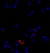
increase in time, but a large increase in information content. Fig.1. Images captured from IN Cell Analyzer 3000. Primary rat
Fluorescent probes Hoechst 33342, Mitotracker deep
hepatocytes following 4 hour exposure to Staurosporine. A:
red (633) and Annexin V (red) were obtained from
composite readout using red (Mitotracker deep red) and blue
Demonstrates use of IN Cell Analyzer in live
Molecular Probes. Draq 5 was obtained from Biostatus
(Hoechst 33342) laser lines and transmitted light (histology stain)images. Highlighted area is shown individually as the (B) blue,
Ltd. Probe mixtures were made up in warmed
(C) red and (D) transmitted light channels respectively. Images
hepatocyte growth media, and dosed in a final volume
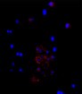
Fig.3. Images captured from IN Cell Analyzer 3000. Primary ratcan be analyzed in real time or offline.
of 100µl for 15 min. NADPH dependent nitro blue
hepatocytes following 4 hour exposure to Staurosporine. A:Control culture stained using Hoechst 33342 (blue) and Annexin
tetrazolium reductase was used as descrbed by
Cells were found to be viable at all doses (Fig. 2), cell
V (red). B: Culture dosed with 1µM Staurosporine, stained usingMurphy et al. (Methods in Cell Science, 21:31-38.1999)
number dropping to approximately 80% of control
Hoechst 33342 (blue) and Annexin V (red).
values at the highest dose. The three measurements
IN Cell assays have increased sensitivity whencompared to traditional assays e.g. Alamar Blue.
Amersham Biosciences Limited 2002 - All rights reserved. Amersham Biosciences. Amersham Place Little Chalfont Buckinghamshire England U.K. HP7 9NA. Amersham
Amersham and Amersham Biosciences are trademarks of Amersham plc.
This poster was presented at the 8 annual Conference of the Society of Biomolecular
Biosciences AB SE-751 84 Uppsala Sweden. Amersham Biosciences Corp 800 Centennial Avenue PO Box 1327 Piscataway NJ 08855 USA. Amersham Biosciences Europe GmbH,
All goods and services are sold subject to terms and conditions of sale of the company within the Amersham Biosciences
Screening, 22-26 September 2002. The Hague, Netherlands.
Munzinger Strasse 9, D-79111, Freiburg, Germany.
group which supplies them. A copy of these terms and conditions are available on request. * To whom all correspondence should be addressed.
P.O. Box 70168 Springfield, OR 97475 Examples of Eligible and Ineligible Expenses The following expenses are commonly requested for reimbursement from Flexible Spending Accounts (FSAs). This list is not comprehensive and is subject to change. In order for any expense to be eligible under your FSA, supporting documentation from your healthcare provider is required. Documentation must inclu
DELAWARE HEALTH AND SOCIAL SERVICES _______________________________________________________________ Division of Public Health _______________________________________________________________ Delaware AIDS Drug Assistance Program (ADAP) Formulary as of September 27, 2011 Antiretrovirals Abacavir (Ziagen) Abacavir/lamivudine (Epzicom) Amprenavir (Agenerase) Atazanavir (Reyataz) C


 High Throughput Multiplexed Cellular Toxicity