Jan-olov.bovin.nu

rearrangement is initiated by the presence of a heteroatom J ˆ 0.8, 2.2 Hz, 1H), 6.50 (d, J ˆ 12.4 Hz, 1 H), 6.17 (dd, J ˆ 1.3, and there must be other reactions similar to this one.
12.4 Hz, 1H), 4.97 ± 4.93 (m, 2 H), 1.18 (s, 9H); 13C NMR (50 MHz, CDCl3): d ˆ 153.9, 153.7, 145.0, 132.3, 129.9, 129.8, 127.2, 125.5, 121.4, 111.3, 110.7, 106.6, 35.9, 29.3. 17: 1H NMR (300 MHz, CDCl3): d ˆ 7.57 (d, J ˆ 2.2 Hz, 1 H), 7.50 (s, 1H), 7.38 (d, J ˆ 8.5 Hz, 1 H), 7.17 ± 7.29 (m, German version: Angew. Chem. 1999, 111, 2753 ± 2755 4H), 7.09 (d, J ˆ 8.3 Hz, 2H), 6.69 (d, J ˆ 2.2 Hz, 1H), 6.49 (d, J ˆ 12.3 Hz, 1H), 6.07 (d, J ˆ 12.3 Hz, 1 H), 5.09 (s, 1H), 4.96 (s, 1H), 3.40 Keywords: heterocycles ´ isomerizations ´ photochemistry ´ (s, 2 H); 13C NMR (75 MHz, CDCl3): d ˆ 154.1, 145.2, 145.1, 139.4, 132.4, 130.5, 130.3, 129.0, 128.2, 127.3, 126.1, 125.3, 121.3, 116.6, 110.8, 106.6, 42.5. 18: 1H NMR (300 MHz, CDCl3): d ˆ 7.61 ± 7.58 (m, 2H), 7.42 (d, J ˆ 8.5 Hz, 1H), 7.28 (td, J ˆ 2.1, 8.5 Hz, 1 H), 6.73 (dd, J ˆ 0.9, [1] H. E. Zimmerman in Rearrangements in Ground and Excited States, 2.1 Hz, 1H), 6.64 (d, J ˆ 12.6 Hz, 1 H), 5.93 (dd, J ˆ 12.6, 26.2 Hz, 1H), Vol. 3 (Ed.: P. de Mayo), Academic Press, New York, 1980, pp. 131 ± 4.75 (ddd, J ˆ 1.2, 2.7, 16.4 Hz, 1H), 4.56 (dd, J ˆ 2.7, 47.4 Hz, 1 H). (E)- 19: 1H NMR (300 MHz, CDCl3): d ˆ 7.66 (s, 1H), 7.61 (d, J ˆ 2.1 Hz, [2] a) G. Kaupp, Angew. Chem. 1980, 92, 245 ± 277; Angew. Chem. Int. Ed.
1H), 7.46 (d, J ˆ 8.7 Hz, 1 H), 7.41 (dd, J ˆ 1.5, 8.7 Hz, 1H), 7.09 (d, J ˆ Engl. 1980, 19, 243 ± 275; b) J. Saltiel, J. L. Charlton in Rearrangements 15.3 Hz, 1H), 6.80 (d, J ˆ 15.3 Hz, 1 H), 6.75 (d, J ˆ 2.1 Hz, 1 H), 5.46 in Ground and Excited States, Vol. 3 (Ed.: P. de Mayo), Academic (s, 1 H), 5.42 (s, 1 H); 13C NMR (75 MHz, CDCl3): d ˆ 155.1, 145.7, Press, New York, 1980, pp. 25 ± 89; c) D. H. Waldeck, Chem. Rev. 1991, 138.8, 133.7, 131.1, 127.9, 124.5, 123.4, 120.0, 115.2, 111.7, 106.7.
91, 415 ± 436; d) G. S. Hammond, J. Saltiel, A. A. Lamola, N. J. Turro, [8] R. E. Kellogg, W. T. Simpson, J. Am. Chem. Soc. 1965, 87, 4230 ± 4234.
J. S. Bradsham, D. O. Cowan, R. C. Counsell, V. Vogt, C. Dalton, J.
[9] R. B. Woodward, R. Hoffmann, The Conservation of Orbital Symme- Am. Chem. Soc. 1964, 86, 3197 ± 3217; e) G. S. Hammond, N. J. Turro, try, Academic Press, New York, 1970.
Science 1963, 142, 1541 ± 1553; f) F. D. Lewis, A. M. Bedell, R. E.
[10] T. D. Doyle, W. R. Benson, N. Filipescu, J. Am. Chem. Soc. 1976, 98, Dykstra, J. E. Elbert, I. R. Gould, S. Farid, J. Am. Chem. Soc. 1990, 112, 8055 ± 8064; g) R. A. Caldwell, J. Am. Chem. Soc. 1970, 92, 1439 ± [11] E. E. Van Tamelen, T. L. Burkoth, R. H. Greeley, J. Am. Chem. Soc.
1441; h) D. Schulte-Frohlinde, H. Blume, H. Gusten, J. Phys. Chem.
1962, 66, 2486 ± 2491; i) H. Gusten, D. Schulte-Frohlinde, Chem. Ber.
[12] 23: 1H NMR (300 MHz, CDCl3): d ˆ 7.39 (d, J ˆ 1.9 Hz, 1H), 7.02 (d, J ˆ 16.2 Hz, 1H), 6.84 (d, J ˆ 16.2 Hz, 1 H), 6.41 (dd, J ˆ 1.9, 3.1 Hz, [3] F. D. Lewis, Acc. Chem. Res. 1979, 12, 152 ± 158.
[4] a) F. B. Mallory, C. W. Mallory, Org. React. 1980, 30, 1; b) F. B.
[13] 24: 1H NMR (300 MHz, CDCl3): d ˆ 7.59 (d, J ˆ 1.9 Hz, 1H), 7.39 (d, Mallory, C. S. Wood, J. T. Gordon, J. Am. Chem. Soc. 1964, 86, 3094 ± J ˆ 8.6 Hz, 1 H), 7.26 (d, J ˆ 8.6 Hz, 1 H), 6.72 (d, J ˆ 1.9 Hz, 1 H), 5.01 3102; c) M. V. Sargent, C. J. Timmons, J. Chem. Soc. 1964, 5544 ± 5552; d) F. B. Mallory, J. T. Gordon, C. S. Wood, J. Am. Chem. Soc. 1963, 85, [14] A. R. Katritzky, L. Serdyuk, L. Xie, J. Chem. Soc. Perkin Trans. 1 1998, 828 ± 829; e) W. M. Moore, D. D. Morgan, F. R. Stermitz, J. Am. Chem.
[15] 26: 1H NMR (300 MHz, CDCl3): d ˆ 7.74 (d, J ˆ 8.3 Hz, 1H), 7.72 (s, [5] a) C. E. Loader, C. J. Timmons, J. Chem. Soc. C 1967, 1677 ± 1681; b) B.
1H), 7.37 (d, J ˆ 5.6 Hz, 1 H), 7.32 (dd, J ˆ 1.6, 8.3 Hz, 1H), 7.25 (d, J ˆ Antelo, L. Castedo, J. Delamano, A. GoÂmez, C. LoÂpez, G. Tojo, J. Org.
5.6 Hz, 1H), 6.52 (d, J ˆ 12.3 Hz, 1 H), 6.19 (d, J ˆ 12.3 Hz, 1H), 5.03 Chem. 1996, 61, 1188 ± 1189; c) G. Karminski-Zamola, L. FisÏer-Jakic, (s, 1 H), 4.98(s, 1H), 1.71 (s, 3H); 13C NMR (75 MHz, CDCl3): d ˆ K. Jakopcic, Tetrahedron 1982, 38, 1329 ± 1335; d) K. Oda, H. Tsujita, 142.0, 139.4, 138.2, 134.1, 132.7, 129.3, 126.5, 125.4, 123.8, 123.7, 121.7, M. Sakai, M. Machida, Heterocycles 1996, 42, 121 ± 124; e) G.
Karminski-Zamola, M. Bajic, Synth. Commun. 1989, 19, 1325 ± 1333.
[6] The photochemical reaction was carried out in degassed dichloro- methane solution in a Pyrex tube using a Rayonet reactor (350 nm) at room temperature. The product 12 was isolated by column chroma- tography on silica gel, no other by-products were observed in the crude 1H NMR spectra. 12: 1H NMR (200 MHz, CDCl3): d ˆ 7.59 (d, J ˆ 2.2 Hz, 1H), 7.52 (d, J ˆ 0.8 Hz, 1H), 7.40 (d, J ˆ 8.5 Hz, 1H), 7.27 Cryo-TEM Snapshots of Ferritin Adsorbed on (dd, J ˆ 1.7, 8.5 Hz, 1H), 6.72 (dd, J ˆ 0.8, 2.2 Hz, 1 H), 6.53 (d, J ˆ 12.2 Hz, 1H), 6.17 (d, J ˆ 12.2 Hz, 1 H), 5.02 ± 4.97 (m, 2H), 1.70 (s, 3H); 13C NMR (50 MHz, CDCl3): d ˆ 154.0, 145.2, 142.1, 132.7, 132.1, Daniel Klint, Gunnel Karlsson, and Jan-Olov Bovin* 129.5, 127.1, 125.4, 121.3, 116.9, 110.6, 106.6, 22.1; MS (70 eV, EI): m/z (%): 184 (67, [M‡]), 169 (100, [M‡ À CH3]), 155 (59), 141 (89), 115 The development of cryo techniques in combination with (39), 105 (41), 91 (26), 77 (26); HR-MS: calcd for C13H12O: 184.0888; transmission electron microscopy (TEM) has increased the number of biological systems that can be studied.[1] Cryo- [7] Spectral data for compound 13: 1H NMR (200 MHz, CDCl3): d ˆ 7.58 ± 7.57 (m, 2H), 7.39 (d, J ˆ 8.5 Hz, 1H), 7.32 (dd, J ˆ 1.6, 8.5 Hz, TEM has become a common tool for the investigation of 1H), 6.71 (d, J ˆ 2.2 Hz, 1 H), 6.50 (d, J ˆ 12.3 Hz, 1 H), 6.10 (d, J ˆ water/surfactant systems[2] and nucleation of inorganic crys- 12.3 Hz, 1H), 4.98 (s, 2H), 2.10 (q, J ˆ 7.4 Hz, 2H), 1.01 (t, J ˆ 7.4 Hz, tals in solution.[3,4] We present for the first time direct imaging 3H); 13C NMR (50 MHz, CDCl3): d ˆ 154.0, 147.7, 145.1, 132.6, 131.2, 129.7, 127.2, 125.4, 121.3, 113.5, 110.8, 106.6, 28.8, 12.8. 14: 1H NMR 3): d ˆ 7.63 (s, 1 H), 7.58 (d, J ˆ 2.2 Hz, 1 H), 7.38 (s, 2H), 6.71 (d, J ˆ 2.2, Hz, 1H), 6.50 (d, J ˆ 12.4 Hz, 1H), 6.07 (d, J ˆ National Center for HREM, Inorganic Chemistry 2 12.4 Hz, 1H), 4.95 (s, 2 H), 2.41 (sept, J ˆ 6.8 Hz, 1 H), 1.12 (d, J ˆ Center for Chemistry and Chemical Engineering 132.4, 130.4, 130.0, 127.3, 125.4, 121.3, 111.5, 110.8, 106.6, 34.0, 21.7. 15: 3): d ˆ 7.59 ± 7.56 (m, 2 H), 7.39 (d, J ˆ 8.5 Hz, 1 H), 7.33 (dd, J ˆ 1.6, 8.5 Hz, 1H), 6.71 (dd, J ˆ 0.7, 2.2 Hz, 1H), 6.49 (d, J ˆ 12.3 Hz, 1 H), 6.07 (d, J ˆ 12.3 Hz, 1 H), 5.00 ± 4.96 (m, 2 H), 2.08 (t, J ˆ 7.6 Hz, 2 H), 1.54 ± 1.35 (m, 2H), 0.83 (t, J ˆ 7.4 Hz, 3): d ˆ 154.0, 146.0, 145.1, 132.5, 131.1, 129.7, 129.5, 127.2, 125.4, 121.3, 114.7, 110.7, 106.6, 38.2, 21.5, 13.8. 16: [**] The Swedish Natural Science Research Council is acknowledged for 1H NMR (200 MHz, CDCl3): d ˆ 7.66 (m, 1H), 7.55 (d, J ˆ 2.2 Hz, financial support. The Knut and Alice Wallenberg Foundation is 1H), 7.45 (dd, J ˆ 1.8, 8.6 Hz, 1 H), 7.35 (d, J ˆ 8.6 Hz, 1H), 6.64 (dd, acknowledged for funding the equipment at the Biomicroscopy Unit.
 WILEY-VCH Verlag GmbH, D-69451 Weinheim, 1999 of solutions containing proteins interacting with zeolite Y 
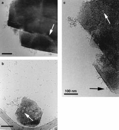
crystals. Pronounced adsorption of ferritin on ultrastable zeolite Y crystals is shown to be correlated to protein aggregation. Adsorption of ferritin molecules on low- and high-silica zeolite Y crystals results in different arrangements of the protein molecules. It is also shown that structural information, like unit cell parameters, can be obtained from inorganic materials present in vitrified solutions.
In recent years the use of zeolites, crystalline aluminum silicates, has been an alternative or complement to common biochemical methods in the purification of proteins.[5,6] Earlier studies of the adsorption of proteins on ultrastable zeolite Y (USY) have shown the protein adsorption to be dependent on pH value and ionic strength.[5] The process is believed to be predominantly mediated by protein aggregates interacting Ferritin is an iron storage protein present in animals and plants. The protein is spherical with a diameter of approx- imately 12 nm. The iron core can consist of up to 4500 Fe ions, thus giving a useful contrast when viewed with the micro- scope. In TEM images ferritin molecules are identified as black spots; the iron core is about 5 nm in diameter. The high contrast from the iron core makes it difficult to see the protein shell. ApoferritinÐthat is, ferritin without an iron coreÐis Figure 1. TEM images of frozen aqueous solutions containing horse spleen identified as a doughnut-shaped molecule with a diameter of ferritin and USY crystals. The lighter areas of various shapes on the crystallites are interpreted as mesopores formed during dealumination; 12 nm. Generally, a protein shows a minimum solubility some are indicated by white arrows. Ferritin molecules are identified as around its isoelectric point (IEP), the pH value in solution black spots approximately 5 nm in diameter. a) Adsorption of ferritin on where the sum of charges on the protein is zero. The IEP for USY in a 20 mmolLÀ1 glycine solution at pH 3.0. Ferritin aggregates of ferritin is pH 4.5, and solutions with pH values at or close to various sizes are adsorbed in a patchlike manner on {111} surfaces of the twin crystal. The image has been subjected to background subtraction the IEP will contain protein aggregates. The adsorbent matrix (script in DigitalMicrograph software) in order to increase the contrast.
is USY, derived from the parent structure NaY by postsyn- b) USY and ferritin in a 20 mmolLÀ1 glycine solution at pH 3.0 also thetic dealumination.[7] Both zeolites have the same FAU containing 150 mmolLÀ1 NaCl. Ferritin molecules are mainly present as structure, but differ in Si/Al ratio and surface texture. The Si/ monomers and a large number of small aggregates consisting of five Al ratio is 2.6 in NaY and 230 in USY, indicating a great molecules or less. Only a few ferritin molecules are adsorbed on the zeolite.
Poorly defined surfaces of the crystallite are probably also due to the difference in framework charge density and thus in surface dealumination. c) Ferritin aggregates of various sizes adsorbed on a USY charge density. The textural difference is due to mesopore crystal in 20 mmolLÀ1 buffer solution at pH 5.2 containing 150 mmolLÀ1 formation during dealumination,[8] and results in less defined NaCl. Aggregates of various sizes were adsorbed on crystals. The black arrow indicates a dimer on the {100} surface of the crystal (see text for Partial precipitation of ferritin was obtained in 20 mmolLÀ1 acetate solution at pH 3.6 (low ionic strength) and also in 20 mmolLÀ1 acetate at pH 5.2 containing 150 mmolLÀ1 NaCl ionic strength) resembled the distribution observed at pH 3.0 (high ionic strength). No visible precipitation was observed in and at high ionic strength (Figure 1b). Also in this case, there solutions of 20 mmolLÀ1 glycin at pH 3.0, either with or was a high degree of monomers and small aggregates (3 ± without the addition of 150 mmolLÀ1 NaCl, or in 20 mmolLÀ1 10 molecules) and only a minor interaction with the zeolite acetate at pH 5.2. Solutions of low ionic strength at pH 3.0 crystals. The solution at pH 5.2 with 150 mmolLÀ1 NaCl was showed the presence of a wide distribution of intermediate- clarified by centrifugation prior to the incubation with USY in sized protein aggregates mainly consisting of less than 15 ± 20 order to remove large precipitates. The majority of the wide ferritin molecules. Cryo-TEM images of such solutions also distribution of protein aggregates remaining in solution containing USY showed that the aggregates were mainly consisted of less than 30 ferritin molecules. In the presence adsorbed on the zeolite crystals (Figure 1 a). Note that the of these aggregates the adsorption on USY crystals was again solution surrounding the crystallite is depleted of ferritin more pronounced (Figure 1c). As in the case shown in molecules. There is no indication of monolayer adsorption, Figure 1a aggregates of various sizes are adsorbed, with the but rather a patchlike distribution of protein aggregates; only solution surrounding the crystallite being depleted of ferritin.
a fraction of the adsorbed molecules interacts directly with the In Figures 1a ± c the presence of mesopores in the USY adsorbent surface. Addition of 150 mmolLÀ1 salt at pH 3.0 crystals can be observed as lighter areas of various shapes, increased the solubility of ferritin by reducing the average aggregate size. The increase in protein monomer concentra- Adsorption of ferritin molecules on NaY and USY crystals tion was followed by a decrease in the adsorbed amounts of was performed in 20 mmolLÀ1 acetate solutions at pH 3.6.
ferritin (Figure 1b). The aggregate distribution at pH 5.2 (low Again, the solution consisted of a wide distribution of mainly  WILEY-VCH Verlag GmbH, D-69451 Weinheim, 1999 
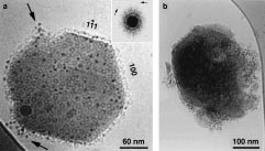
large protein aggregates, some containing 50 ± 100 molecules.
consequently low in the pH 3.0 solution containing Nevertheless, the adsorption of ferritin resulted in a different 150 mmolLÀ1 NaCl (Figure 1b.) Increasing the pH value protein arrangement on the NaY crystals than on the USY leads to an increased deprotonation of the USY crystal crystals. In Figure 2 a the NaY crystal is viewed approximately surfaces and hence an increase in negative charges. The reduced adsorption in 20 mmolLÀ1 buffer solution at pH 5.2 (low ionic strength) is reminiscent of the case of the solution at pH 3.0 containing 150 mmolLÀ1 NaCl (Figure 1b) and is explained by a cooperative effect of two different events: 1) The net negative charge on the ferritin molecules is enough to maintain a low degree of aggregation, and 2) as the protein and the USY surfaces have charges of the same sign, interaction is reduced due to repulsive forces. The addition of salt causes a shielding effect of the repulsive forces between the USY surfaces and ferritin molecules, enabling protein aggregates to be formed and adsorbed (Figure 1c).
The different adsorption behavior of ferritin on NaY and USY (Figure 2) could be explained by the great difference in chemical composition. On the NaY surface approximately Figure 2. TEM images after the adsorption of ferritin molecules on NaY every third tetrahedron contains Al, and it therefore displays and USY crystals in a 20 mmolLÀ1 acetate buffer solution at pH 3.6. a) The a higher negative electric potential. As adsorption was NaY crystal is viewed approximately along [110], as indicated by the power spectrum in the inset; the arrows in the inset indicate the reciprocal performed in a solution of low ionic strength and at a pH distance to the {111} reflections. Due to partial precipitation of ferritin, the value below the IEP for ferritin, the interactions are much solution contained a wide distribution of mainly large aggregates (50 ± more ionic in character at the NaY crystal surfaces than at 100 molecules). Nevertheless, ferritin molecules are almost uniformly distributed on the NaY crystal, except for a few aggregates indicated by arrows. The diameter of the doughnut shaped molecules is 12 nm, corresponding to the size of apoferritin. b) The adsorbed ferritin molecules on USY result in a different pattern. Large aggregates are adsorbed in a patchlike manner only covering parts of the crystallite. The crystallite in (b) Suspensions of ultrastable zeolite Y particles (USY-HSZ-390HOA, Tosoh has approximately the same orientation as that in (a).
Co., Japan) in 20 mmolLÀ1 buffer solutions, either with or without the addition of 150 mmolLÀ1 NaCl, were degassed and ultrasonicated prior to sedimentation. Supernatants containing suitable particle sizes were ob- along [110], displaying the {111} and {100} surfaces. The tained by centrifugation at 1000 ± 1500 g for 5 min. Diameters of the zeolite reciprocal distances to the {111} reflections correspond to crystals thus collected were in the range of 150 ± 1000 nm, as estimated from 1.42 nm in real space, which agrees well with the calculated low-magnification cryo-TEM images. Adsorption was performed by incubation of horse spleen ferritin with zeolite supernatant suspensions distance of 1.42 nm for NaY (Fd3Åm, a0 ˆ 2.47 nm). There does for 1 h on a rocking table at room temperature (ferritin concentration not seem to be any preference for adsorption on any approximately 0.02 mg mLÀ1). Sample were prepared by applying a drop of particular crystallographic surface. The ferritin molecules solution (8 mL) onto a lacey, carbon film covered TEM copper grid in a are uniformly distributed on the NaY crystal. The molecules temperature and humidity controlled environment vitrification system adsorbed on the surfaces projected perpendicular to the (CEVS).[9] The excess solution was blotted off, and the remaining liquid film was plunged into a reservoir of liquid ethane at its freezing point ( À viewing direction show traces of hexagonal close packing. A 1838C). The vitrified specimen was transferred by an Oxford CT3500 cryo- relatively large amount of molecules displays a doughnut holder into a Philips CM120 BioTwin Cryo; the temperature was never appearance. Since many of these molecules are at the same allowed to raise above À 160 8C. The images were recorded under low-dose focus as the dark spots, it can be ruled out that the doughnut conditions using 117-kV energy filtered electrons on a Gatan 791 cooled shape is due to focal effects. Therefore, the NaY crystal is covered with both ferritin and apoferritin molecules. On USY ferritin is adsorbed patch-wise as aggregates, and only a German version: Angew. Chem. 1999, 111, 2736 ± 2738 fraction of the molecules interacts directly with the surface (Figure 2b). The crystal in Figure 2b also displays distinct Keywords: electron microscopy ´ proteins ´ zeolites crystallographic surfaces, although they are partly damaged [1] J. Dubochet, M. Adrian, J.-J. Chang, J.-C. Homo, J. Lepault, A. W.
Ferritin molecules carry a net positive charge at pH values McDowall, P. Schultz, Q. Rev. Biophys. 1988, 21, 129 ± 228.
below the IEP. As deprotonation of terminal hydroxyl groups [2] P. K. Vinson, J. R. Bellare, H. T. Davis, W. G. Miller, L. E. Scriven, J.
occurs on the USY surfaces, attractive electrostatic forces Colloid Interface Sci. 1991, 142, 74 ± 91.
[3] O. Regev, Langmuir 1996, 12, 4940 ± 4944.
arise between the zeolite surfaces and the ferritin molecules.
[4] M. T. Kennedy, B. A. Korgel, H. G. Monbouquette, J. A. Zasadzinski, These electrostatic interactions may play a role in the Chem. Mater. 1998, 10, 2116 ± 2119.
adsorption of the ferritin aggregates at ionic strength below [5] D. Klint, H. Eriksson, Protein Expression Purif. 1997, 10, 247 ± 255.
pH 4.5. However, addition of salt breaks up protein ± protein [6] Y. C. Yu, Y. C. Huang, T. Y Lee, Biotechnol. Prog. 1998, 14, 332 ± 337.
interactions and at the same time causes a shielding effect by [7] J. Scherzer, ACS Symp. Ser. 1984, 248, 157 ± 200.
[8] J. Lynch, F. Raatz, P. Dufresne, Zeolites 1987, 7, 333 ± 340.
decreasing the Debye length, thus reducing the effect of the [9] J. R. Bellare, H. T. Davis, L. E. Scriven, Y. Talmon, J. Electron Microsc.
electrostatic forces. The extent of adsorption of ferritin was  WILEY-VCH Verlag GmbH, D-69451 Weinheim, 1999

Source: http://www.jan-olov.bovin.nu/pdfs/paper/P95.pdf

Microsoft word - anaesthetic inguinal hernia pre-op info sheet1.doc

Anaesthetic Information Sheet for Hernia Surgery Overall philosophy: We aim to provide you with the best and safest anaesthetic care available anywhere. We aim to achieve the best possible pain relief. We aim to do what we can to make your stay in hospital as pleasant as possible. We aim to treat you the way you want to be treated: We do not “do” mediocre. This sheet lists what we do for the

Microsoft word - prescriptiondrugcardsprs.doc

147 Shore Drive • Somerville, Massachusetts 02145 (617) 666-5958 • edgar@consumerworld.org DRUG DISCOUNT CARDS: A PRESCRIPTION FOR SAVINGS But Comparing Card Discounts is a Bitter Pill (BOSTON- JULY 23) -- Consumers without prescription drug coverage can save money using little-known drug discount cards, especially free ones, according to a price comparison survey releas

Copyright © 2010-2014 Metabolize Drugs Pdf