Tadalafil entfaltet seine Wirkung über eine selektive Hemmung der PDE5, wodurch die Konzentration von cGMP im glatten Muskelgewebe stabil bleibt. Diese biochemische Modulation resultiert in einer langanhaltenden Relaxation der Gefäßwände. Der Wirkstoff wird nach oraler Einnahme effizient resorbiert, mit einer Bioverfügbarkeit von rund 80 %. Seine Halbwertszeit von bis zu 36 Stunden ist innerhalb dieser Substanzklasse außergewöhnlich. Abgebaut wird er in der Leber, hauptsächlich durch CYP3A4, mit anschließender biliärer Exkretion. Typische unerwünschte Wirkungen entstehen durch eine verstärkte Vasodilatation, etwa Kopfschmerzen oder Flush. Pharmakologisch wird cialis generika vor allem durch die verlängerte Wirkungsdauer charakterisiert.
Multi-frequency high-field epr study of iron centers in malarial pigments
Multi-Frequency High-Field EPR Study of Iron Centers in Malarial Pigments
Andrzej Sienkiewicz,§,† J. Krzystek,‡ Bertrand Vileno,† Guillaume Chatain,¶ Aaron J. Kosar,¶
D. Scott Bohle,*,¶ and La´szlo´ Forro´†
Institute of Physics, Polish Academy of Sciences, Al. Lotniko´w 32/46, 02-668 Warsaw, Poland, Institute of Physicsof Complex Matter, EÄcole Polytechnique Fe´de´rale, CH-1015 Lausanne, Switzerland, National High Magnetic FieldLaboratory, Florida State UniVersity, 1800 East Paul Dirac DriVe, Tallahassee, Florida 32310, Department ofChemistry, McGill UniVersity, 801 Sherbrooke Street West, Montreal, Quebec H3A 2K6, Canada
Received January 13, 2006; E-mail: scott.bohle@mcgill.ca
Malaria, whose most severe form is caused by a protozoan
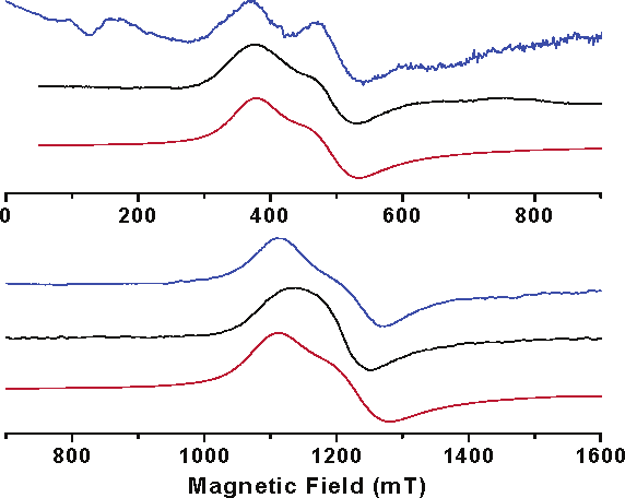
parasite, Plasmodium falciparum (Pf), remains the world’s most prevalent vector-borne disease. The spread of chloroquine-resistant strains (CQR) of Pf and the absence of a suitable replacement for this once effective antimalarial created an urgent need to understand the biochemistry behind the drug’s action.1 The intraerythrocytic growth stage of Pf involves hemoglobin proteolysis as the primary nutrient source with the concomitant release of free heme. The liberated heme is detoxified by the parasite into an inert crystalline material, called malarial pigment, or hemozoin.2,3 According to the recent hypothesis, chloroquine inhibits heme aggregation in ring Figure 1. The low-field (high geff) turning point in the EPR spectra of
or early-stage malaria trophozoites.4 It has been shown that
hemozoin (blue) and -hematin (black) at 34 (top) and 94 GHz (bottom) at
hemozoin is chemically,2,5 spectroscopically,2,3 and crystallographi-
10 K. The red traces are powder-pattern simulations using the following
cally6 identical and isostructural to its synthetic phase, -hematin.
spin Hamiltonian parameters: |D| ) 5.80 cm-1, |E| ) 0.20 cm-1, g⊥ )
The magnetic susceptibility measurements and Mo¨ssbauer spec-
1.90 (top) and |D| ) 5.85 cm-1, |E| ) 0.075 cm-1, g⊥ ) 1.95 (bottom).
-hematin suggested the presence of a single high-
2, which is located 0.47 Å out of the plane of
2) iron environment of largely axial symmetry in its
the porphyrin and forms a relative short bond, 1.889 Å, with one
bulk phase.3,7 Recently, the crystal structure of
of the oxygens of the protoporphyrin-IX propionic acid substit-
solved by X-ray powder diffraction.8 The structure is surprising in
uents.13 The distance between the two Fe ions within the dimer is
that rather than being a coordination polymer, as widely held
9.05(1) Å, with a mean porphyrin plane separation of 4.44 Å. Due
before,2,3 it is a chain of dimers formed by the FeIII-protoporphy-
to their offsets, the nearest atoms in the porphyrin rings are separated
rin-IX molecules through reciprocal iron-carboxylate bonds to
by 5.00(1) Å, but the nearest iron neighbors are in adjacent unit
one of the propionic side chains of each porphyrin. The dimers
cells where the Fe-Fe separations are 7.86(1), 8.04(1), and
then build chains linked by hydrogen bonds in the crystal.8
Despite the congruous nature of much of the spectroscopic data
for the natural and synthetic phases, there is still considerableambiguity in interpretation of the Electron Paramagnetic Resonance(EPR) results concerning the characterization of their local Feenvironment.2,7,9-12 For example, previous work performed atconventional X-band (9.5 GHz) frequency suggested a rhombicsymmetry of the zero-field splitting (zfs) tensor, which was not inagreement with Mo¨ssbauer results.7 In this report, we present thedefinite conclusions of the spin state and properties of the groundstate of hemozoin and
frequency high-field EPR (HFEPR) to simplify and fully interpret
EPR spectra of hemozoin were acquired at cryogenic tempera-
spectra of the ferric ion. At the same time, we will use the HFEPR
tures on two Bruker Elexsys spectrometers equipped with TE011
data to find correlations between structure and magnetic properties
resonators: Q-band (34 GHz) at the EPFL, and W-band (94 GHz)
of both natural and synthetic malarial pigments.
-Hematin was investigated in a wide range of
Malarial pigment (hemozoin) was isolated from a K-1 chloro-
frequencies (27-500 GHz) in transmission-type single-pass spec-
quine-resistant strain of Plasmodium falciparum. The pigment was
trometers at the NHMFL.16 At each frequency, the
isolated from late-stage trophozoites following nonproteolytic
spectrum consists of a strong turning point at high effective geff ∼
methods.14 Homogeneous single-phase microcrystalline powders of
4.3-5.5, depending on frequency, and a weak turning point at geff
-hematin were prepared by anhydrous dehydrohaloge-
2. At low frequencies (27-94 GHz), the high geff absorption
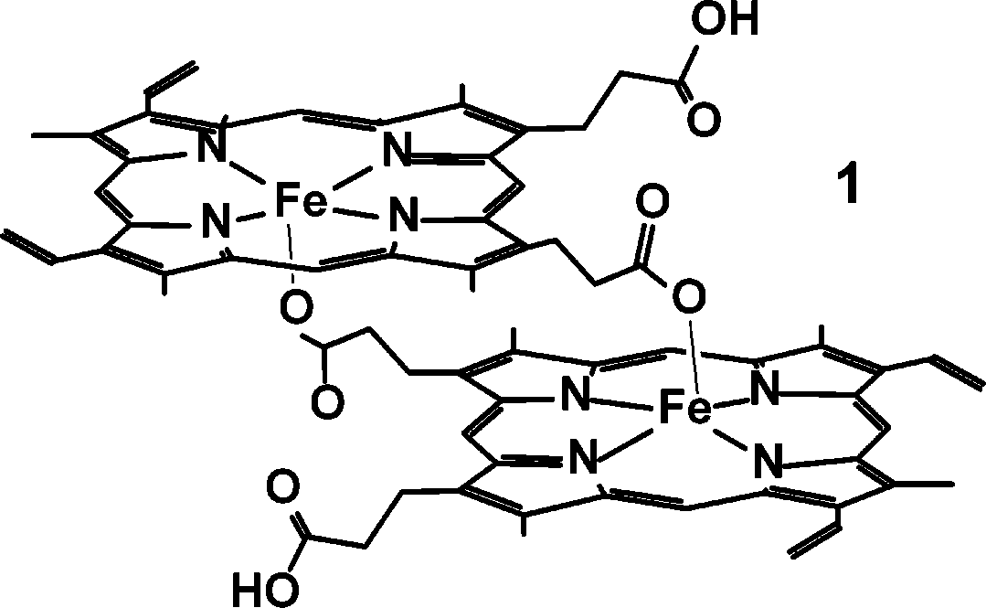
nation of hemin.15 The structure of a building block of the malarial
line is partly split into two components (Figure 1). In this frequency
range, the spectra of hemozoin are nearly identical to those of
2 dimer, 1, has a five-coordinate
-hematin. At higher frequencies, the splitting disappears, and a
single line is observed (Figure S1 in the Supporting Information).
† EÄcole Polytechnique Fe´de´rale de Lausanne. ‡
At high frequencies (above ca. 270 GHz), a new turning point
is detected in the -hematin spectra (Figure S1), appearing at yet
4534 9 J. AM. CHEM. SOC. 2006, 128, 4534-4535 10.1021/ja058420h CCC: $33.50 2006 American Chemical Society C O M M U N I C A T I O N S
troscopy as 7.5 cm-1.19 As for the small but measurable rhombicparameter E, its presence was detected both in Fe(DmePP-IX)-(N3)18 and in the above-mentioned complex with an axial acetategroup,19 and apparently depends on the symmetry of the liganditself. The propionate linker between the two porphyrins in
-hematin may be conducive to producing such a small rhombic
distortion. An explanation of the effect of averaging of the E valuewith increasing EPR frequency remains outside the scope of thepresent work, but we mention here the analogy with averaging the
Figure 2. Resonance field versus quantum energy (or frequency) depen-
dipolar broadening of EPR resonances in solids with increasing
dence of turning points in the powdered sample of -hematin. Experimental
observation frequency in the intermediate exchange regime.20 This
points are marked by squares. Below 100 GHz, the average position of the
analogy seems to be particularly attractive since the averaged
partly split low-field resonance was taken. Lines were simulated using best-
perpendicular turning point at high frequencies actually gets
fitted spin Hamiltonian parameters as in text. Blue lines, parallel turningpoints; red lines, perpendicular turning points. For clarity, only those
narrower with increasing frequency and field in the solid, unlike
transition branches are plotted that are actually observed experimentally.
the situation existing in a magnetically diluted system, such as
The broken line corresponds to the frequency (378.2 GHz) at which the
bottom trace in Figure S1 was recorded. Acknowledgment. This work was partially supported by the
lower fields than the previously described high g
Polish KBN Grant #2-PO3B-090-19, by the IHP-Contracts HPRI-
new signal approaches zero field at ca. 350 GHz and starts
CT-2001-00140, by G1MA-CI-2002-4017 (CEPHEUS) of the
increasing in field again at higher frequencies. Figure 2 summarizes
European Commission (A.S.), by the Burroughs-Wellcome Fund
(P.W.S. and D.S.B.), by a CIHR Traineeship grant (C.G.), and by
the Swiss National Science Foundation (B.V. and L.F.). We thank
The HFEPR spectra obtained for hemozoin and -hematin can
Dr. A. Ozarowski (NHMFL) for his simulation software, and Peter
only be interpreted as originating from high-spin FeIII (S ) 5/
W. Stephens for his valuable assistance.
described by a nearly axial spin Hamiltonian of the standard form,
Supporting Information Available: EPR spectra of -hematin in
comprising both Zeeman and zfs terms, H ) BgS +D[S 2 -
the high-frequency regime at T ) 10 K are shown in Figure S1
(1 page, print PDF). This material is available free of charge via the
Sy ). An observation of the zero-field transition
at 350 GHz, which, in the axial case, is equal to 2|D|, yields the
value of |D| ) 5.83 cm-1. To our best knowledge, this is the first
References
successful EPR detection of an inter-Kramers transition in a heme-
like molecule. A complete set of intrinsic spin Hamiltonian
parameters is delivered through a simultaneous fit to all the observed
(2) Slater, A. F. G.; Swiggard, W. J.; Orton, B. R.; Flitter, W. D.; Goldberg,
D. E.; Cerami, A.; Henderson, G. B. Proc. Natl. Acad. Sci. U.S.A. 1991,
resonances, as previously described:17 D ) +5.85(1) cm-1, E ) 0,
g⊥ ) 1.95(1), g ) 2.00(1) (the actual positive sign of D is given
(3) Bohle, D. S.; Conklin, B. J.; Cox, D.; Madsen, S. K.; Paulson, S.; Stephens,
P. W.; Yee, G. T. In Inorganic and Organometallic Polymers II, AdVanced
by simulations of single-frequency spectra shown in Figure S1). Materials and Intermediates; Wisian-Neilson, P., Allcock, H. R., Wynne,
The splitting in the perpendicular turning point of the intra-Kramers
K. J., Eds.; American Chemical Society: Washington, DC, 1994; pp 497-
transition at low frequencies indicates, however, that the zfs tensor
(4) Dorn, A.; Stoffel, R.; Matile, H.; Bubendorf, A.; Ridley, R. G. Nature
may not be entirely axial, as suggested by HFEPR. A fit to the
1995, 374, 269-271.
Q-band spectra, where the observed splitting is the largest, yields
(5) Fitch, C. D.; Kanjananggulpan, P. J. Biol. Chem. 1987, 262, 15552-
the |E| value of 0.2 cm-1, that is, a rhombicity of the zfs tensor
(6) Bohle, D. S.; Dinnebier, R. E.; Madsen, S. K.; Stephens, P. W. J. Biol.
|E/D| equal to 0.035. This is in very good agreement with the
Chem. 1997, 272, 713-716.
(7) Bohle, D. S.; Debrunner, P.; Jordan, P. A.; Madsen, S. K.; Schultz, C. E.
previous Mo¨ssbauer spectroscopy conclusions.7 The effect of the
J. Am. Chem. Soc. 1998, 120, 8255-8256.
splitting decreasing and finally vanishing at higher frequencies is,
(8) Pagola, S.; Stephens, P. W.; Bohle, D. S.; Kosar, A. D.; Madsen, S. K. Nature 2000, 404, 307-310.
however, puzzling since simulations show that it should increase
(9) Schoffa, G. Nature 1964, 203, 640-641.
rather than decrease. We see spin exchange as a tentative explana-
(10) Arese, P.; Schwarzer, E. Ann. Trop. Med. Parasitol. 1997, 91, 501-516. (11) Cammack, R.; Patil, D. S.; Linstead, D. J. Chem. Soc., Fraday Trans.
tion of this phenomenon (see below). 1994, 90, 3409-3410.
In general, however, our results show that magnetic exchange
(12) Bremard, C.; Girerd, J. J.; Kowalewski, P.; Merlin, J. C.; Moreau, S. Appl.
within each dimer is negligible, even weaker than suggested by
Spectrosc. 1993, 47, 1837-1842.
(13) Bohle, D. S.; Kosar, A. D.; Stephens, P. W. Acta Crystallogr. 2002, D58,
previous susceptibility measurements.3 Otherwise, the ground spin
state of the dimer would be zero or an integer number, and not, as
(14) Ashong, J. O.; Blench, I. P.; Warhurst, D. C. Trans. R. Soc. Trop. Med.Hyg. 1989, 83, 167-172.
observed by us, S ) 5/2. Apparently, the significant distance between
(15) Bohle, D. S.; Helms, J. B. Biochem. Biophys. Res. Commun. 1993, 193,
the two Fe centers and the number of chemical bonds between them
(16) (a) Hassan, A. K.; Pardi, L. A.; Krzystek, J.; Sienkiewicz, A.; Goy, P.;
make magnetic exchange very inefficient.
Rohrer, M.; Brunel, L.-C. J. Magn. Reson. 2000, 142, 300-312. (b)
The spin Hamiltonian parameters obtained for the first time with
Zvyagin, S. A.; Krzystek, J.; van Loosdrecht, P. H. M.; Dhalenne, G.; Revcolevschi, A. Physica B 2004, 346-347, 1-5.
high accuracy for a heme-like system are within the range observed
(17) Krzystek, J.; Zvyagin, S. A.; Ozarowski, A.; Trofimenko, S.; Telser, J. J.
in similar mononuclear Fe centers. It is known that the zfs parameter
Magn. Reson. 2006, 178, 174-183.
(18) Brackett, G. C.; Richards, P. L.; Caughey, W. S. J. Chem. Phys. 1971, D in five-coordinated Fe(III) complexes depends on the nature of
the axial ligand. Thus, D as measured by us is very similar in value
(19) Bominaar, E. L.; Ding, X. Q.; Gismelseed, A.; Bill, E.; Winkler, H.;
to that determined for Fe in protoporphyrin-IX dimethyl ester with
Trautwein, A. X.; Nasri, H.; Fischer, J.; Weiss, R. Inorg. Chem. 1992, 31, 1845-1854.
a fluoride axial ligand (5.0 cm-1) as measured by far-IR magnetic
(20) Krzystek, J.; Sienkiewicz, A.; Pardi, L.; Brunel, L. C. J. Magn. Reson.
spectroscopy.18 A more relevant comparison can be made with
1997, 125, 207-211.
(21) van Kan, P. J. M.; van der Horst, E.; Reijerse, E. J.; van Bentum, P. J.
another porphyrinic Fe(III) complex, with Fe axially ligated by an
M.; Hagen, W. R. J. Chem. Soc., Faraday Trans. 1998, 94, 2975-2978.
acetate residue, whereas D was determined by Mo¨ssbauer spec-
J. AM. CHEM. SOC. 9 VOL. 128, NO. 14, 2006 4535
Information and prices are correct at the time of publication (July 2011), however may be subject to change. *P.O.A – Please phone 1300 552 512 for clarification of the fee. $33.15 (Medicare rebate available under certain circumstances) thromboembolism or First degree relative who has a prove defect of Antithrombin, protein C/S or APCR ADH $30.70 (Invoice from $31.15 (Invoi
CAPITOLATO TECNICO FORNITURA IN SERVICE DI APPARECCHIATURE E METERIALE DI CONSUMO PER CONFORMITA’ ACCREDITAMENTO PER IL SERVIZIO DI IMMUNOEMATOLOGIA E MEDICINA TRASFUSIONALE DEL PRESIDIO OSPEDALIERO “SAN GIOVANNI DI DIO” DI CROTONE PER UN PERIODO DI MESI TRENTASEI LOTTO n° 1: Sacche per la raccolta di sangue intero con accessori in SERVICE Cod. CIG: 526828457C
 Multi-Frequency High-Field EPR Study of Iron Centers in Malarial Pigments
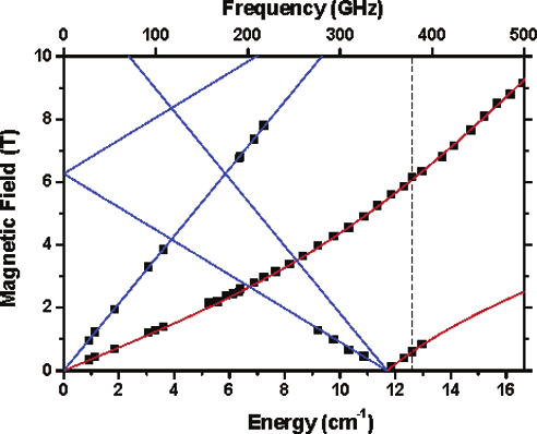 C O M M U N I C A T I O N S